Ultrasound (sonography) is a medical diagnostic technique which allows one to visualize and therefore examine a part of the human anatomy. The process incorporates the use of high frequency sound waves emitted from a probe and directed into a body. These sound waves penetrate and encounter the different tissue interfaces as they travel through the body. When sound encounters tissues or tissue planes, part of the wave is reflected back to the receivers in the same probe. Different tissues interfaces cause a "reflective pattern" which is then sent to a computer. The computer processes the information and produces an image, which is sent to a video screen, printer and or videocassette recorder. These images are then studied either "live" or at a later time to rule out any number of pathologies.
Ultrasonography Imaging
Ultrasound (sonar) has its roots in the military, and in brief, it was used in submarines for detecting the surface and shape of the ocean floor. In medicine, B-mode or B-scan simply means the scanning of a part of the human body. Such a scan is either live, where the image is seen on a vide screen and printed at the time it is acquired, or scanned now and compiled later into an image for study.
An ultrasound waves are sent into the tissues, part of the sound is reflected back to the probe while others continue into deeper tissues. Much of this is dependent on the frequency of the probe, which is being used. Unlike the probes used for physical therapy which are designed for deep heating of tissues, diagnostic ultrasonography probes cause no heating or tissue damage. Ultrasound is safe during pregnancy. Ultrasound uses no radiation unlike x-rays, Cat Scans and other such modalities.
Scanning Podiatric Pathology
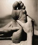
An advantage of diagnostic ultrasound besides its safety, is the ability to perform live active and/or passive range of motion studies. The ability to move a particular part of the anatomy, observe and record motion studies such as partial or complete tendon, muscle, and ligament tears should not be understated. To enable the physician to diagnose cases, record, and document these studies increases the likelihood of better treatment and end result. Please note that MRI, CAT scans, and x-rays all do not allow for motion studies. Although diagnostic ultrasound is not intended to eliminate those studies, one should realize that the cost of an ultrasound is about one-tenth that of an MRI. Should the physician receive satisfactory information from the musculoskeletal ultrasound and avoid the need of an MRI scan, the patient and/or insurance companies benefit in those savings.
Podiatric Diagnostic Ultrasound